Wednesday, April 25, 2007

most recent publications

A few things that some of you might be interested in:

1. Plasmacytoid Urothelial Carcinoma of the Urinary Bladder, Report of Seven Cases. K. T. Mai, P. C. Park, H. M. Yazdi, E. Saltel, S. Erdogan, W. A. Stinson, I. Cagiannos, C. Morash. Case Study of the Month, European J Urology 50 (2006) 1111-1114.

2. The 3-Dimensional Structure of Isolated and Small Foci of Prostatic Adenocarcinoma, The Morphologic Relationship Between Prostatic Adenocarcinoma and Prostatic Intraepithelial Neoplasia. K. T. Mai, B. F. Burns, W. A. Stinson, C. Morash. Research Article, Appl. Immunohistochem Mol Morphol 15(1) 2007: 50-55.

And something a little older:

3. Proposed technique for sectioning of mastectomy specimens and submission of tissue for microscopic examination of breast carcinoma. K. T. Mai, H. M. Yazdi, B. F. Burns, D. G. Perkins, D. Mirsky. Correspondence, Histopathology 39, 2001: 323-327.
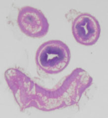
Spectrum Webviewer

With Quorum Technologies' able assistance, I'm going to try to allow access to a web-based version of Aperio Technologies' software called Spectrum Webviewer - simply by clicking on the image. The images that I have been posting are far too small to appreciate anything even remotely subtle. This will enable manipulation of the images, zooming in and out, etc. Wait 'till you see it! It's pretty neat.

Much ado

You may notice that a couple of my previous posts have been edited and re-posted.

I regret that my comments may have caused some consternation.

Sunday, April 22, 2007

New Image

The images illustrate an interesting case that I saw recently. This young male patient had a long history of recurrent pancreatitis of unknown etiology. He was otherwise healthy. A Whipple's procedure was performed and we found an area of thickening in the wall of the duodenum adjacent to the Ampulla of Vater (gross image) which turned out to be ectopic pancreas (micro image). Notice in the gross image that the pancreatic tissue is between the mucosa and serosa. The bile and pancreatic ducts were markedly ectatic and the pancreas was diffusely fibrotic.

Sunday, April 01, 2007

Professional Extenders?

Many of you have no doubt read with interest the one-page article in the Winter, 2007 CAP Newsletter entitled Professional Extenders for Pathologists, authored by Dr. Raymond Maung. Dr. Maung states that he intends to discuss this issue at the CAP AGM in June. There are many points within the article with which I could take issue but in the interests of brevity and diplomacy, I will make just one comment: there are so many pathologists across this country whose practice depends upon PAs that I think raising this discussion at the CAP will be enlightening.
It is refreshing and encouraging that an attempt is being made to measure the portion of the pathologists' workload traditionally dedicated to grossing. If the numbers are anywhere near accurate, they will further validate the practice of PAs. I support the thinking that up to 1/3 of a pathologist's time at a tertiary-care facility is, or ought to be, dedicated to grossing. PAs and Histotechs who perform gross descriptions have never been given the recognition that they deserve. All provincial ministries have been essentially unaware, for over 30 years, that someone other than pathologists have been doing all of this work.
I am so glad to see that the CAP has included a day-long session specifically for PAs at this year's meeting. I regret that I will likely not be able to attend but want to send my best wishes to all those who will be there.